stages of mitosis under microscope Mitosis meiosis lab observing tips onion root cell division vs tip slides cycle mitotic getty reschke ed evolution biology leerlo
Mitosis is a fascinating process that plays a crucial role in the growth and development of living organisms. It is a tightly regulated sequence of events that ensures the accurate distribution of genetic material to new cells. Let’s delve into the stages of mitosis and explore the wonders of cell division!
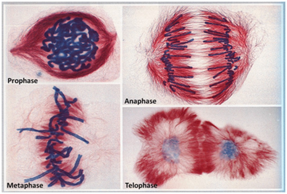
Prophase:
In the first stage of mitosis, prophase, the chromatin condenses and becomes visible as distinct chromosomes under a microscope. Each chromosome consists of two identical sister chromatids held together by a structure called the centromere. The nuclear envelope starts to break down, and the centrosomes move to opposite poles of the cell, forming spindle fibers that will later help separate the chromosomes into new cells.
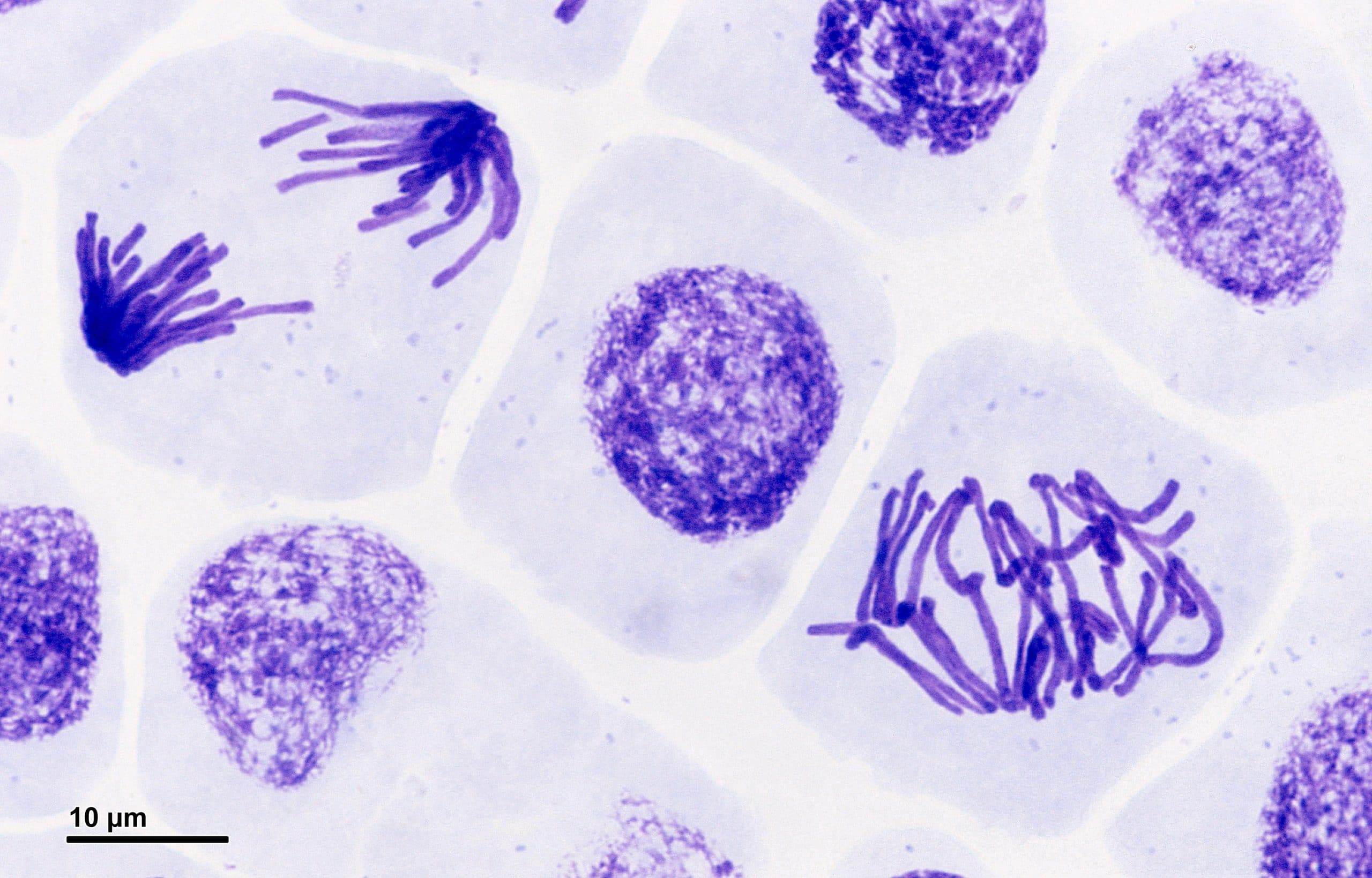 This stunning image above showcases the beauty of prophase in a pressed root meristem of Vicia faba cells. The vibrant colors and distinct chromosomes captivate the eye, demonstrating the intricate nature of cell division.
Metaphase:
During metaphase, the chromosomes align themselves along the equator of the cell, known as the metaphase plate. The spindle fibers attach to the centromeres of each chromosome, ensuring that they are properly positioned for separation. This alignment is crucial for the equal distribution of genetic material to the new cells.
 Above is a mesmerizing image showcasing metaphase under a microscope. The symmetrical arrangement of the chromosomes along the metaphase plate is truly a sight to behold. It reminds us of the incredible precision and orchestration taking place within our cells.
Anaphase:
As anaphase unfolds, the centromeres holding the sister chromatids together split, allowing the spindle fibers to pull the chromatids towards opposite poles of the cell. This ensures that each new cell will receive an identical set of chromosomes. The cell starts elongating as the poles move apart.
Mitosis is a crucial process responsible for growth, tissue repair, and asexual reproduction in various organisms. Understanding the stages of mitosis provides us with a deeper appreciation for the complexity and beauty of life at the cellular level.
So next time you look under a microscope and observe the incredible dance of chromosomes during mitosis, take a moment to marvel at the wonders of nature. The intricate choreography of cell division is truly a masterpiece!
If you are looking for Major Functions of Mitosis and Meiosis | hubpages you’ve visit to the right web. We have 5 Images about Major Functions of Mitosis and Meiosis | hubpages like Mitosis - Stages - Prophase - Metaphase - TeachMePhysiology, Stages Of Mitosis And Meiosis Under Microscope - Micropedia and also Stages Of Mitosis And Meiosis Under Microscope - Micropedia. Here it is:
Major Functions Of Mitosis And Meiosis | Hubpages
 hubpages.commitosis cell meiosis cells stages different division functions microscope under anaphase stage steps biology function between major difference reproduction organisms
Stages Of Mitosis And Meiosis Under Microscope - Micropedia
microspedia.blogspot.commitosis microscope cells prophase interphase metaphase anaphase meiosis telophase mitose microbiology ทยา วว
Top Tips For Observing Mitosis Lab
/139812087-56a2b3cd3df78cf77278f2cb.jpg) www.thoughtco.commitosis meiosis lab observing tips onion root cell division vs tip slides cycle mitotic getty reschke ed evolution biology leerlo
www.thoughtco.commitosis meiosis lab observing tips onion root cell division vs tip slides cycle mitotic getty reschke ed evolution biology leerlo
Microscope Pictures Of Mitosis Stages - Micropedia
 microspedia.blogspot.commitosis microscope phases cells meiosis labeled division cytokinesis cycle telophase pmat cas miamioh
Mitosis - Stages - Prophase - Metaphase - TeachMePhysiology
teachmephysiology.commitosis prophase cell cells anaphase root stages meristem microscope vicia faba phases metaphase cycle division pressed celular figure microscopio meiosis
Microscope pictures of mitosis stages. Top tips for observing mitosis lab. Major functions of mitosis and meiosis